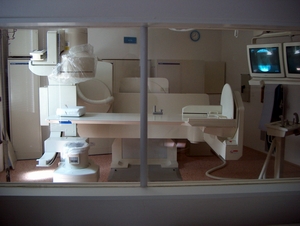
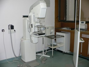
There are skiagraphic and fluoroscophic examinations performed at the Clinic of Radiology 1st. MF UC Na Bulovce. At the present times there are available 3 digital skiascopic-skiagraphic walls, 2 second category walls and one 1st category wall for examining oesophagus, stomach, small intestine and colon and for other examinations as fistulography, phlebography, hysterosalpingography (HSG) and perimyeolography (PMG).
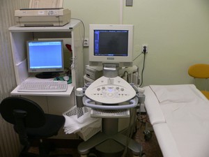
Ultrasound examination is possible by using four available ultrasound machines and is aimed specially at diagnostics of abdomen. But we perform many more examinations such as the examination of soft tissue for orthopaedic purposes, the examination of thyroid gland with puncture possible, the biopsy-examinations under ultrasound control in abdominal area and the examination of mammary gland also with possible puncture. There are Doppler´s blood vessels examinations performed in neck area and in lower extremities.
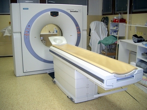
Nowadays there is available one of the most modern MDCT gears in the world made by Siemens (Sensation 40) with 40 raws of detectors and with 0,37s lamp rotation. It allows to perform common examination of every part of the body with high degree of quality and also to perform even more specialised examinations such as CT angiography of brain, of carotids, abdomen and lower extremities and examination of pulmonary embolism as well. This machine is capable of 3D reconstruction of all human organs- including skeleton. Thanks to very advanced software it is possible to perform also virtual and (CAD) examinations as virtual CT endoscopy and virtual CT colonoscopy. Also CT perfusion examination is possible- urgent CT perfusion to diagnose ischemia and also examinations of perfusion of parenchymatic organs and tumours. In pulmonary diagnostics it is used to identify nodules of lung parenchyma. It is also possible to determine quantitative amount of calcium in skeleton by using densitometry with minimal irradiation doses.
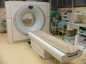
There is still available and functioning another CT gear with 16 rows of detectors (Siemens Sensation 16)- it is used for common examination and also in cooperation with the Clinic of Oncology for planning and for radiation fields´ location for radiotherapy.
The focus of the clinic is centred on interventional methods- vascular and nonvascular. Vascular interventional methods are performed such as percutaneous transluminal angioplastics and stents of peripheral arteries (PTA), embolisation of tumours and vascular malformation of CEA (a. carotis ext.) and chemoembolisation of tumours in abdomen, in retroperitoneum and in pelvis and trombolysis of peripheral arteries.
Nonvasular interventional examinations are performed such as biopsies under CT and US control, abscess- drainage of whole body, drainage of pseudocysts in chronic pancreatitis, also drainage of fluid collection in acute severe pancratitis, also percutaneous drainage of biliary tract, laser nucleotomy of intervertebral discs under sciascopic control, radiofrequent interstitial ablation of meta process in liver nad sceleton, alcoholisation of sympathetic nerve system and cysts, adhesing of cavities in abdomen and pelvis by using tissue glue. Another example of interventional examinations with high degree of specialisation are stenting of oesophageal tumor- stenosis, dilatation of oesophagus and small intestine and dilatation of rectosigmatal stenosis.
Since 2004 has been established mamographic screening- mammography is performed by modern mammographic gear Senovision GE with a possibility of stereobiopsy of focus in mammary gland by using digital stereotaxy. Supplementary method is sonography with a possibility of immediate mammar gland-biopsy under ultrasonic guidance.
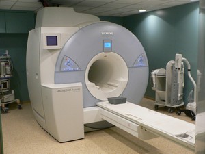
There is also available the most modern magnetic resonance workplace - Avanto made by Siemens, that allows to operate with data from several coils´elements at the same time. This gear performes examination of every part of a human body in the shortest time possible, and it belong among the most quiet ones. The possible specialised examinations available are MR angiography, MR cholangiography and pancreaticography, MR enteroclysis, MR of mammary gland, that specifies the character of a pathological focus, the biopsy is also possible ( at the present time the only one in Czech Republic), there are also functional MR examinations- MR spectroscopy and MR perfusion.
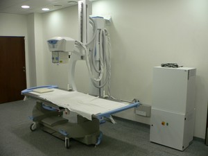
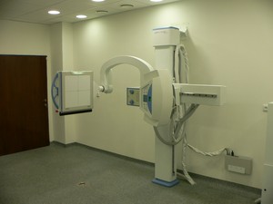
As a digitalisation process continues, we have started to use undirect digitiser KODAK CR500 and since 2006 also new digitiser DR by Canon, that skiagraphs to flat panel by using C-arm. This technology is much easier for radiographers to use, it decreases the dosage, improves patients´ comfort and improves also the quality of images.